Protein Synthesis
Protein Synthesis
DNA contains a series of nitrogenous bases. These nitrogenous bases code the sequence of amino acids that are to be linked together to form a particular protein.
This series of nitrogenous bases is called a gene.
Thus, DNA contains genes. One gene code for one protein.
Three nitrogenous bases code one amino acid. They are called a codon. They code the primary structure of the protein which folds to form its tertiary structure. This tertiary structure is important for the function of proteins which serve as enzymes for different metabolic reactions.
For example:
There are 20 different types of amino acids.
As there are only 4 bases, 43=64. Thus, there are 64 possible pairings of the nitrogenous base. Amino acids are coded by more than one triplet.
For example AAA and AAG both codes for phenylalanine.
Mechanism Of Protein Synthesis
Proteins are synthesized in the cytosol by ribosomes. The whole process is divided into two steps:
Transcription
• To make protein the copy of genetic code is to be sent to the ribosomes in the form of mRNA this process is called transcription.
Process
• The DNA is unzipped to expose nitrogenous bases.
There are four nucleotides in the cytoplasm(A, U, T, C). They attach with their complementary
• DNA bases.
• The pairing takes place in the following order:
AàU
CàG
GàC
TàA
• When the RNA nucleotides attached with their complementary bases RNA polymerase binds them together. The RNA nucleotides linked together are called messenger RNA. Messenger RNA contains a copy of genetic code which can then be transferred to the ribosomes for making proteins.
Translation
Making of protein from messenger RNA is called translation.
• The mRNA formed breaks from the nucleus and is moved to the cytoplasm.
• The cytoplasm has 20 different amino acids and tRNA.
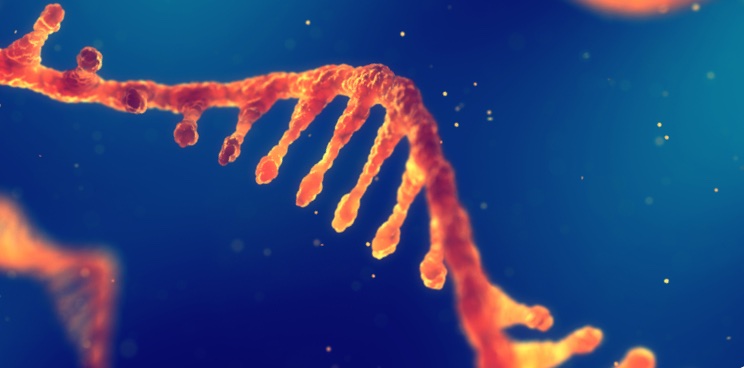
• tRNA is a type of RNA. It consists of an exposed nitrogenous base on one side and amino acid is attached to its other side. The nitrogenous base on tRNA is called an anticodon. The amino acid attached to tRNA is complementary to the anticodon. For example, tRNA with anticodon UAC will have methionine attached to it.
• mRNA will be attached to the ribosome. The first two codons will be attached to the groove in the ribosome. tRNA will bring the anticodon that is complementary to the codon. The amino acids that are attached to the adjacent tRNA will form a peptide bond. mRNA will move within the ribosome bringing in the next codon.
• The first codon is always a start codon that is AUG. it codes for methionine.
• This process will continue until a stop codon is reached.
• A stop codon, for example, UAG does not code any amino acid. Thus, the process of translation is terminated over here.
Gene Mutation
Any abnormal change in the DNA is called a mutation.
These mutations or changes can arise anywhere in the DNA. The cell contains various repair mechanism thus most of the time these mutations are repaired. But, there are times when they persist. This results in a change in the expression of the gene.
Types Of Mutation
Different types of mutations arise in a DNA
Deletion
• A pair of nitrogenous bases can get deleted in a gene.
Insertion
• Nitrogenous bases can be added in gene.
Translocation
• A portion of one chromosome can be translocation to another chromosome making a fusion gene.
Inversion
The sequence of nitrogenous bases is inverted to create a mirror image of the gene.
Result Of Mutations
The change in the sequence of nitrogenous bases results in the formation of an abnormal protein. This is because the sequence of amino acids is changed resulting in a change in the structure of the protein. As the structure is linked to its function thus most of the proteins cannot perform their function.
Sickle Cell Disease
Hemoglobin contains 4 chains of proteins linked together. There are two alpha-globin and two beta-globin as previously said.
In sickle cell disease, there is a mutation in the Beta globin. There is a change in only one base that is it contains T in place of A.
This results in the substitution of valine in place of glutamic acid in the beta chain. The resulting hemoglobin is called HbS.
Normal Hb is called HbA.
Effect Of Mutation
Glutamic acid is hydrophilic it interacts with the aquatic environment to make the Hb molecule stable.
Valine is hydrophobic. Thus, it is insoluble and makes ppts of hemoglobin.
In the case of hypoxia, the HbS stick to each other and make long insoluble fibers. These fibers distort the shape of hemoglobin and make it Sickle-shaped. This sickle-shaped hemoglobin is rigid and cannot pass through small capillaries. This results in hemolysis.
